
What to Know About Breast Density, Your Risk Assessment, and Callbacks.
You made time for your mammogram, an important step for your breast health. Then your results mention dense breast tissue, include a risk score you may not recognize, or you receive a call asking you to return for additional images. What does that mean for you?
Understanding what your results are telling you, and why additional imaging may sometimes be recommended, can help you feel more informed about what comes next.
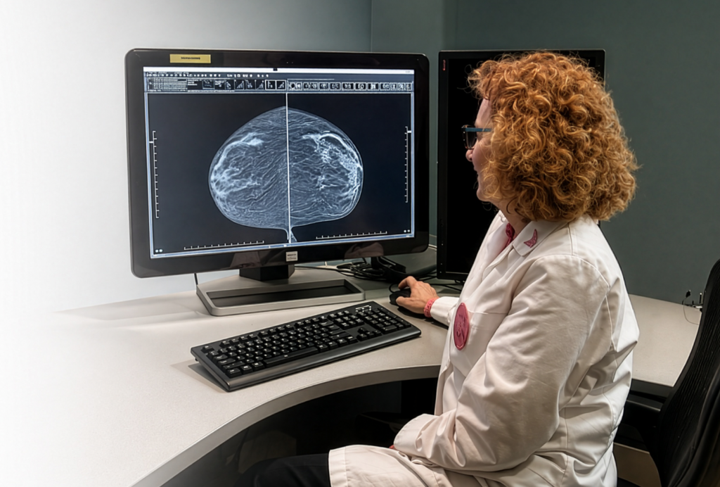
What Does It Mean to Have Dense Breasts?
Dense breasts have a relatively high amount of glandular and fibrous tissue compared with fatty tissue. Having dense breast tissue is common. All breasts contain a combination of fatty tissue and dense breast tissue. On a mammogram, fatty tissue appears darker and is easier to see through, while dense tissue appears more opaque. This can make some cancers more difficult to see within dense tissue.
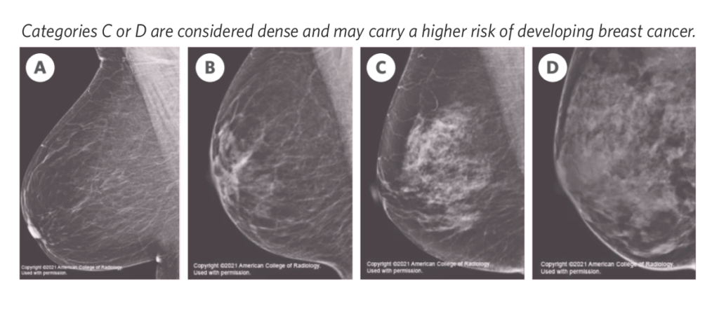
Your breast density is determined by your radiologist based on your mammogram and falls into one of four categories:
A). Almost entirely fatty: The breast is almost entirely fatty tissue
B). Scattered fibroglandular density: The breast is mostly fatty tissue, with some dense areas
C). Heterogeneously dense: The breast is mostly dense tissue, with some fatty areas
D). Extremely dense: Nearly all of the breast is dense tissue, with very little fat
Categories C and D are considered dense breasts. Having dense breast tissue does not mean you have cancer, but it may carry a higher risk of developing breast cancer and can make cancer more difficult to see on a mammogram.
Jefferson Radiology uses 3D mammography, also called breast tomosynthesis, for screening mammograms. It captures multiple low-dose X-ray images from different angles and creates detailed layers of breast tissue for your radiologist to view. This layer-by-layer view can be especially helpful when evaluating dense breast tissue, where overlapping tissue can make abnormalities more difficult to see. Even with 3D mammography, additional imaging may be recommended based on your breast density, overall risk, or a finding that needs a closer look.
If I Have Dense Breasts, Do I Need Additional Imaging?
Depending on your breast density and individual risk factors, additional screening with breast ultrasound or breast MRI may be recommended alongside your mammogram.
_______________________________________________________________
When a screening breast ultrasound may be recommended
A screening breast ultrasound may be recommended alongside a mammogram for patients with dense breast tissue. It may improve the chances of detecting cancer, while mammography remains the gold standard for early detection of breast cancer.
_______________________________________________________________
When a breast MRI may be recommended
Breast MRI may be recommended as an additional screening exam for some patients at higher risk for breast cancer. It does not replace your mammogram. It uses a magnetic field and radio waves to produce detailed images of breast tissue.
_______________________________________________________________
What should I do if I have dense breasts?
If your mammogram shows that you have dense breast tissue, talk with your healthcare provider about whether additional screening may be appropriate for you. Screening breast ultrasound and breast MRI require a referral from your healthcare provider, so they can help you consider your breast density along with your other risk factors and determine the screening plan that’s right for you.
Your breast density is only one part of your overall breast health. Your individual risk factors can also help you and your healthcare provider understand what type of screening may be appropriate for you.
What Does My Breast Cancer Risk Assessment Score Mean?
Your breast cancer risk assessment score estimates your lifetime risk of developing breast cancer.
Jefferson Radiology includes this assessment with every mammogram report shared with you and your referring physician. We use the Tyrer-Cuzick Model, also known as the International Breast Intervention Study (IBIS), to calculate this score based on information collected during your mammogram appointment.
The assessment considers several factors, including your:
- Age
- Breast density
- Personal history of breast biopsies and their results
- Personal history of ovarian cancer
- Family history of breast and ovarian cancer
- Reproductive history, including age of first period or menopause
How do I understand my risk score percentage?
Your risk score percentage reflects your estimated lifetime chance of developing breast cancer. It falls into one of three categories:
- Under 15%: average risk
- 15-19%: intermediate risk
- 20% or higher: high risk
One important thing to remember: your risk score is not a diagnosis. It does not mean you have breast cancer or that you will develop it. Instead, it helps identify factors that may increase your lifetime risk and gives you and your healthcare provider more information to help guide your screening plan. No matter your risk level, your risk assessment does not replace your annual screening mammogram. Depending on your individual risk and breast density, your healthcare provider may recommend additional screening, such as breast ultrasound or breast MRI.
Why Am I Being Called Back After My Mammogram?
A callback means your breast imaging radiologist needs more information or additional images to fully evaluate an area. A call asking you to return for more images can understandably raise questions. You may be asked to return because something looks different from a previous mammogram or because an area needs to be seen more clearly. Most findings that require additional imaging are benign, or noncancerous.
According to the American Cancer Society, fewer than 1 in 10 women who are called back after a mammogram receive a cancer diagnosis. Even so, completing the recommended follow-up is important to find out what a particular finding means for you.
Follow-up often includes a diagnostic mammogram, which takes more detailed views of the area, and sometimes a targeted breast ultrasound.
What happens after I get additional images?
After reviewing your additional images, your radiologist may recommend routine screening, earlier follow-up imaging, or a biopsy, depending on the findings.
- Returning to your regular screening schedule.
- Having follow-up imaging sooner to check whether an area changes.
- Having a biopsy to collect a small tissue sample for examination.
If a biopsy is recommended, imaging helps your radiologist locate the area to sample. Jefferson Radiology offers ultrasound-guided breast biopsy and stereotactic breast biopsy, which use imaging to guide the needle to the area of concern. A pathologist then examines the tissue to determine a diagnosis.
The important thing is to complete the recommended follow-up so your care team has the information needed to determine what comes next.
Questions About Your Mammogram Results?
If you have questions about your breast density, risk assessment, or recommended follow-up, talk with your healthcare provider about what your results mean for you, and whether additional screening may be appropriate. Every mammogram and every patient is different. Our Jefferson Radiology breast imaging team can also help answer questions about recommended imaging and what to expect.
If you’re due for your annual screening, this Breast Cancer Awareness Month booking a mammogram is your Power Move.
___________________________________________________________
REVIEWED FOR CLINICAL ACCURACY
This article has been reviewed by a Jefferson Radiology mammography modality manager to help ensure the medical information is accurate, current, and aligned with today’s imaging practices.
Jennifer G., RT(R)(M)
Registered Technologist Certified in Radiography and Mammography
Last reviewed: September 25, 2026


